Publications
Madan H, Berlot R, Ray NJ, Pernus F, Spiclin Z.
IEEE Journal of Biomedical Health Informatics, 2019 Oct 2
doi: 10.1109/JBHI.2019.2945077
Hennadii Madan, Franjo Pernuš, Žiga Špiclin
Statistical Methods in Medical Research, 2019 Jul;28(7):2196-2209.
doi: 10.1177/0962280217754231
Žiga Lesjak, Alfiia Galimzianova, Aleš Koren, Matej Lukin, Franjo Pernuš, Boštjan Likar, Žiga Špiclin
Neuroinformatics, 2018 Jan;16(1):51-63.
doi: 10.1007/s12021-017-9348-7
Domen Ravnik, Tim Jerman, Franjo Pernuš, Boštjan Likar, Žiga Špiclin
Proceedings Volume 10574, Medical Imaging 2018: Image Processing; 105741J (2018)
doi: 10.1117/12.2293702
Alfiia Galimzianova, Žiga Lesjak, Daniel L. Rubin, Boštjan Likar, Franjo Pernuš, Žiga Špiclin
Journal of Medical Imaging, vol. 5, no. 1, pp. 011007, 2011
doi: 10.1117/1.JMI.5.1.011007
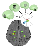
Hennadii Madan, Franjo Pernuš, Žiga Špiclin
Medical Image Computing and Computer Assisted Intervention (MICCAI), 2017
doi: 10.1007/978-3-319-66185-8_86
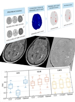
Hennadii Madan, Rok Berlot, Nicola J Ray, Franjo Pernuš and Žiga Špiclin
Predictive Intelligence in Medicine (PRIME), MICCAI, 2018
Žiga Lesjak, Franjo Pernuš, Boštjan Likar, Žiga Špiclin
Neuroinformatics. 2016 Oct;14(4):403-20
doi: 10.1007/s12021-016-9301-1
Alfiia Galimzianova, Franjo Pernuš, Boštjan Likar, Žiga Špiclin
Neuroimage. 2016 Jan 1;124(Pt A):1031-1043
doi: 10.1016/j.neuroimage.2015.09.047
Hennadii Madan, Lina Savšek, Saša Šega Jazbec, Žiga Špiclin
Multiple Sclerosis Journal, vol. 23 (S3), pp. 593-594, 2017
doi: 10.1177/1352458517731286, poster ID: P1133
Lina Savšek, Vojko Strojnik, Žiga Špiclin, Saša Šega Jazbec
Multiple Sclerosis Journal, vol. 23 (S3), p. 747, 2017
doi: 10.1177/1352458517731286, poster ID: EP1417
Lina Savšek, Vojko Strojnik, Alojz Ihan, Žiga Špiclin, Saša Šega Jazbec
Multiple Sclerosis Journal, vol. 23 (S3), p. 974, 2017
doi: 10.1177/1352458517731286, poster ID: EP1849
Hennadii Madan, Franjo Pernuš, Žiga Špiclin
Medical Image Computing and Computer-Assisted Intervention - MICCAI,
Lecture Notes in Computer Science, vol. 10434, pp. 763-771, Springer, 2017
doi: 10.1007/978-3-319-66185-8_86
Datasets
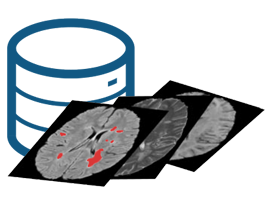
MRI studies of 30 Multiple Sclerosis patients with consensus white matter lesion segmentations performed on a 3D FLAIR, 2D T1-weighted and 2D T2-weighted images.

MRI studies of 30 Multiple Sclerosis patients with consensus white matter lesion segmentations performed on a 3D FLAIR, 2D T1-weighted and 2D T2-weighted images.

MRI studies of 30 Multiple Sclerosis patients with consensus white matter lesion segmentations performed on a 3D FLAIR, 2D T1-weighted and 2D T2-weighted images.

MRI studies of 30 Multiple Sclerosis patients with consensus white matter lesion segmentations performed on a 3D FLAIR, 2D T1-weighted and 2D T2-weighted images.

MRI studies of 30 Multiple Sclerosis patients with consensus white matter lesion segmentations performed on a 3D FLAIR, 2D T1-weighted and 2D T2-weighted images.

MRI studies of 30 Multiple Sclerosis patients with consensus white matter lesion segmentations performed on a 3D FLAIR, 2D T1-weighted and 2D T2-weighted images.
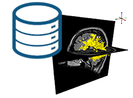
20 Multiple Sclerosis patients with 2 MR studies consisting of T1-weighted, T2-weighted and FLAIR images and white matter lesion changes ground truth segmentation.

20 Multiple Sclerosis patients with 2 MR studies consisting of T1-weighted, T2-weighted and FLAIR images and white matter lesion changes ground truth segmentation.

20 Multiple Sclerosis patients with 2 MR studies consisting of T1-weighted, T2-weighted and FLAIR images and white matter lesion changes ground truth segmentation.

20 Multiple Sclerosis patients with 2 MR studies consisting of T1-weighted, T2-weighted and FLAIR images and white matter lesion changes ground truth segmentation.
Tools

BrainSeg3D is a free volume (3D image) viewer and segmentation tool based on Seg3D. The main goal of BrainSeg3D is to make segmentation of volumes easier, faster and more accurate by providing tools for semi-automated segmentation combined with a user friendly graphic interface.

BrainSeg3D is a free volume (3D image) viewer and segmentation tool based on Seg3D. The main goal of BrainSeg3D is to make segmentation of volumes easier, faster and more accurate by providing tools for semi-automated segmentation combined with a user friendly graphic interface.